Software & Methods
Iterative Immunofluorescence
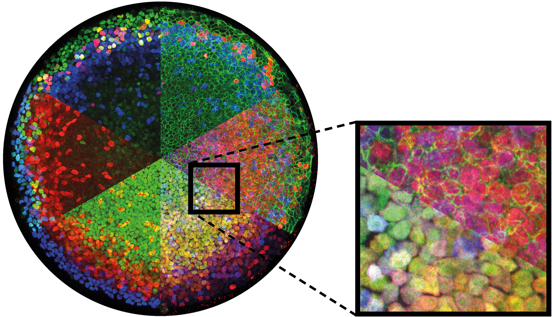
The number of antibody stains that can be performed on a single sample often limits the questions we can ask about how protein levels are related in space at the single-cell level. To address this we optimized the 4i iterative immunofluorescence protocol for 2D human gastruloids and established a computational pipeline to process the data. Find our protocol and processing pipeline for iterative IF here.
Tissue Cartography with ImSAnE
Advances in microscopy have enabled the study of multiscale dynamic problems such as cell tracking across entire embryos. However, increased data size makes storage, processing, and transfer difficult and expensive — most severely for specimens with curved surfaces that do not align with the microscope, where the desired information occupies only a small fraction of the recorded region. Laminar data also pose conceptual problems: even when it is feasible to process data in three dimensions, interpretation in orthographic projections or cross sections is challenging.
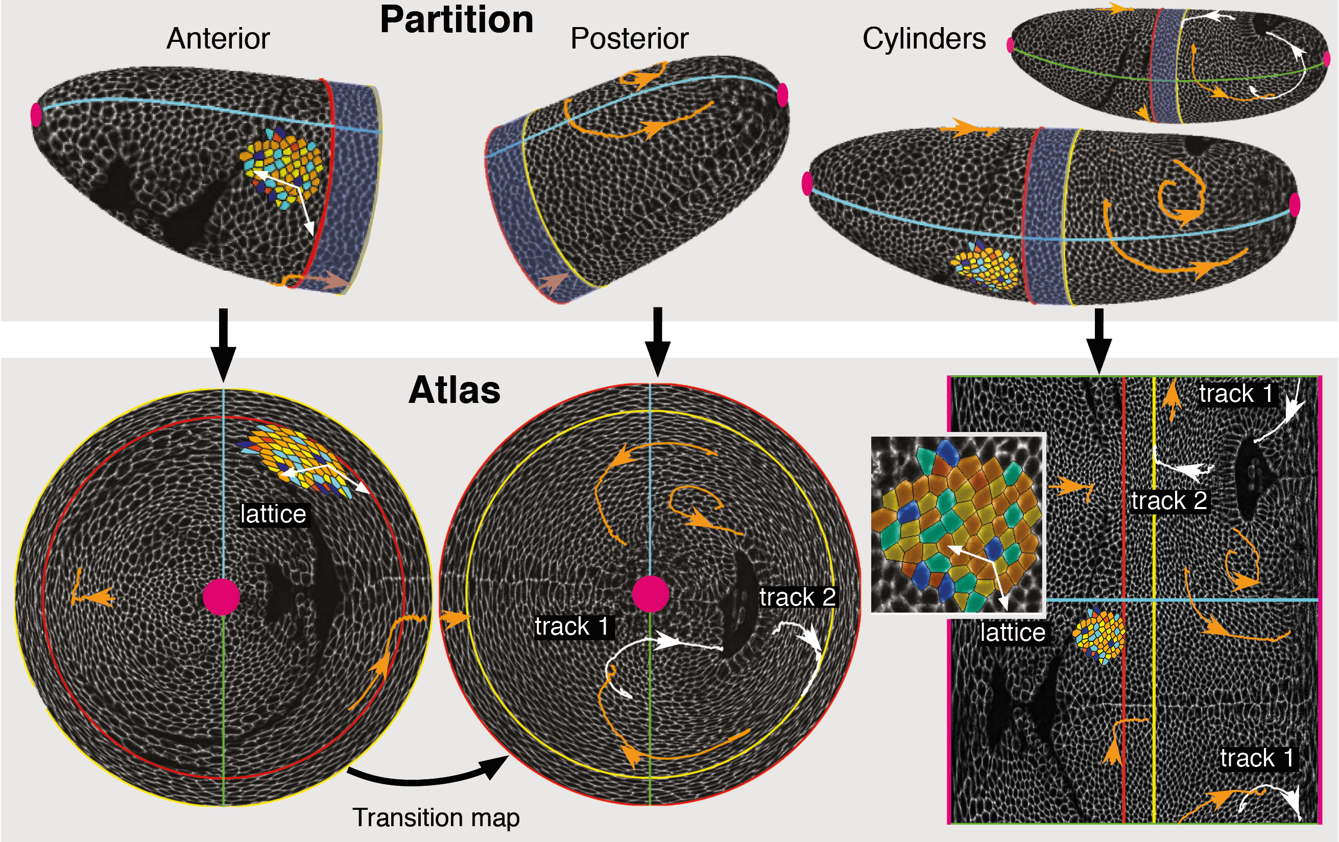
For layered samples, these problems can be solved by considering a surface of interest (SOI) instead of a region of interest and moving into the tissue frame by organizing the data in terms of surface coordinates. This reduces the data from 3D to 2D, analogous to making maps of Earth on a flat grid of longitude and latitude. With Sebastian Streichan, I developed a general framework for tissue cartography using differential geometry, and a practical tool that implements it. Our open-source MATLAB toolbox ImSAnE (Image Surface Analysis Environment) maps surfaces of interest to the plane, stores the geometric metadata required for faithful measurements, and builds an atlas of overlapping maps to create a global picture. It has been tested on a wide range of data, including in-toto analysis of Drosophila gastrulation, tracking cells on a beating zebrafish heart, visualization of zebrafish epiboly, segmentation of epithelial organoids, and nuclear segmentation in mouse endoderm. Read about it in the Nature Methods paper; updated code is at github.com/idse/imsane.
Vertex Model Simulations
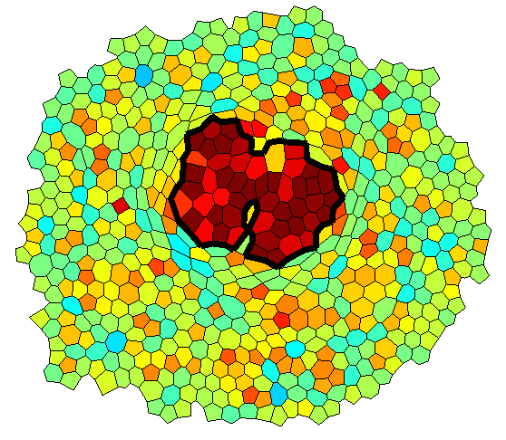
Epithelial monolayers can be approximated by two-dimensional polygonal tilings described by their vertex coordinates. The mechanics of the tissue can then be modeled by assigning tensions to interfaces and pressures to cells, possibly based on levels of cytoskeletal proteins. This framework includes cell rearrangement through T1 transitions and naturally simplifies cell signaling to involve only cells and cell interfaces. I developed a simulation framework that we applied to overgrowing clones in an epithelial monolayer with mechanical feedback on myosin levels. Read the paper that includes the simulations, or download the code at github.com/idse/mechanicalFeedback.